What is Papilledema?
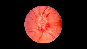
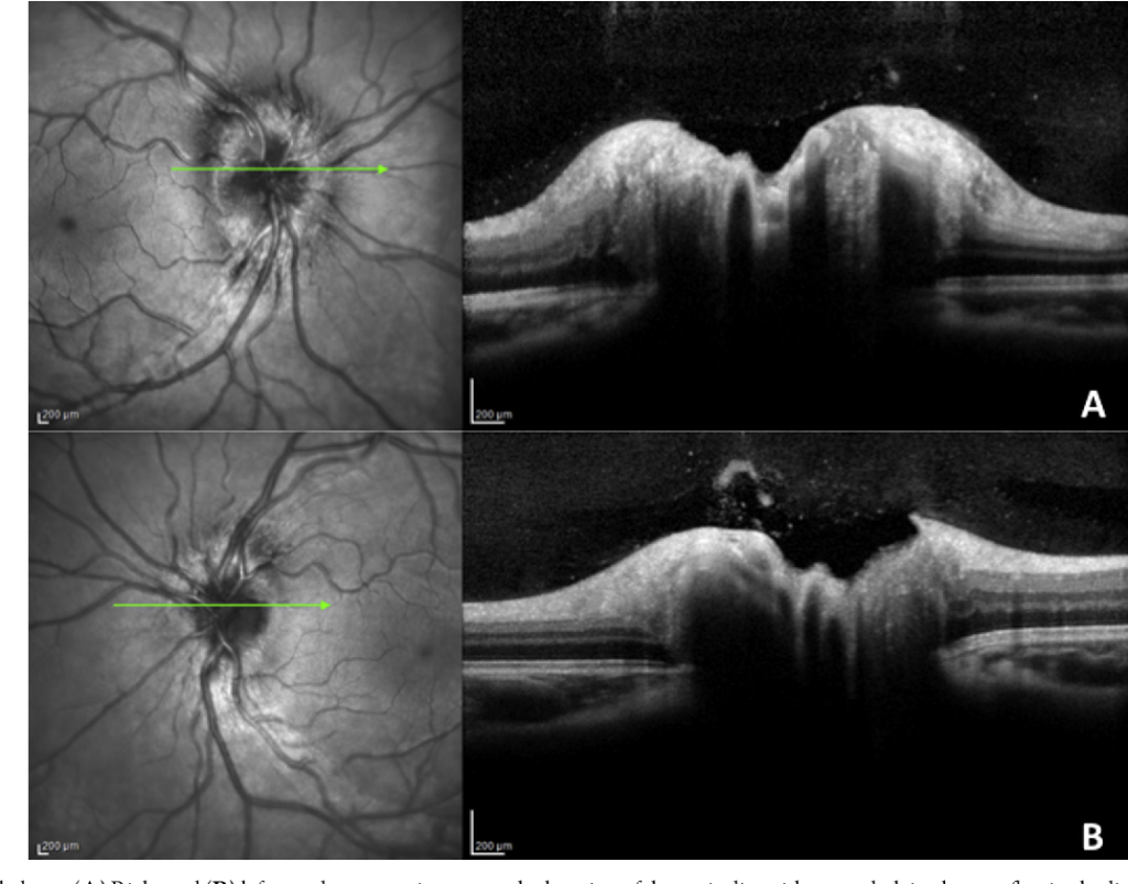
Papilledema is the swelling of the optic disc (the point where the optic nerve enters the eye) due to increased pressure inside the skull, also known as intracranial pressure. This condition is a sign of an underlying problem in the brain, such as a brain tumor, hydrocephalus (excess fluid in the brain), or increased pressure from other causes. Papilledema can lead to vision problems and, if left untreated, may cause permanent damage to the optic nerve and loss of vision.
Unlike optic neuritis, which is inflammation of the optic nerve, papilledema is caused by the pressure that affects the optic disc. It can be identified through an eye examination, where an eye doctor (optometrist or ophthalmologist) will look for signs of swelling at the back of the eye.
What Causes Papilledema?
Papilledema occurs when there is increased pressure inside the skull, leading to a backup of fluid into the optic nerve. This pressure interferes with the normal function of the optic nerve, causing swelling. Several conditions can lead to increased intracranial pressure, including:
1. Brain Tumors
A mass or tumor in the brain can obstruct the normal flow of cerebrospinal fluid (CSF), leading to an increase in pressure inside the skull.
2. Hydrocephalus
Hydrocephalus is a condition where there is an abnormal accumulation of cerebrospinal fluid in the ventricles of the brain, leading to increased pressure.
3. Intracranial Hemorrhage
A bleeding event in the brain, such as a stroke or head injury, can lead to increased intracranial pressure, causing papilledema.
4. Idiopathic Intracranial Hypertension (IIH)
Also known as pseudotumor cerebri, this condition is characterized by increased pressure inside the skull without an obvious cause, such as a tumor or infection.
5. Infections
Certain infections that affect the brain, such as meningitis or encephalitis, can lead to inflammation and increased intracranial pressure, causing papilledema.
6. Venous Sinus Thrombosis
This condition occurs when there is a blood clot in the venous sinuses of the brain, which impedes the normal flow of blood and increases intracranial pressure.
7. Traumatic Brain Injury (TBI)
Head injuries that cause bleeding or swelling in the brain can increase pressure inside the skull and result in papilledema.
What Are the Symptoms?
Papilledema often presents with no noticeable symptoms at first, and it is commonly discovered during an eye exam. However, as the condition progresses, the following symptoms may develop:
1. Vision Problems
- Blurry vision – One of the earliest symptoms of papilledema.
- Temporary vision loss – Often in the form of transient episodes of vision loss that can occur suddenly.
- Double vision (diplopia) – Difficulty seeing clearly, sometimes with overlapping or duplicated images.
2. Headache
Headaches are a common symptom of conditions that lead to increased intracranial pressure. In papilledema, the headache may worsen with time.
3. Nausea and Vomiting
Increased intracranial pressure often causes nausea and vomiting, which may be associated with headaches.
4. Visual Disturbances
People with papilledema may experience visual obscurations (a temporary loss of vision), especially when changing positions (e.g., moving from lying down to standing up).
5. Papilledema-Related Visual Impairment
If the swelling of the optic disc continues and is not treated, the individual may experience permanent vision damage and even blindness.
How is Papilledema Diagnosed?
The diagnosis of papilledema involves several steps:
1. Eye Examination
- Fundoscopy (ophthalmoscopy): A healthcare provider will examine the back of the eye using a special instrument to look for signs of swelling or changes in the optic disc. Papilledema causes the optic disc to appear swollen and blurred.
2. Neurological Examination
- A neurologist may conduct a thorough neurological examination to look for other signs of increased intracranial pressure, such as weakness, altered consciousness, or problems with coordination.
3. Imaging Studies
- MRI or CT scan: These imaging tests are used to look for structural abnormalities in the brain, such as tumors, hydrocephalus, or brain hemorrhages, that could be causing the increased intracranial pressure.
4. Lumbar Puncture (Spinal Tap)
- In some cases, a lumbar puncture may be performed to measure the pressure of the cerebrospinal fluid (CSF) around the brain and spinal cord. This can help diagnose conditions like idiopathic intracranial hypertension (IIH).
5. Visual Field Testing
- This test helps assess any visual impairment caused by papilledema and checks for changes in the patient’s peripheral vision.
How is Papilledema Treated?
Treatment for papilledema focuses on addressing the underlying cause of the increased intracranial pressure. The specific treatment plan will vary depending on the condition causing the papilledema.
1. Treating the Underlying Cause
- If the papilledema is due to a brain tumor, treatment may involve surgery, radiation, or chemotherapy.
- If the condition is caused by hydrocephalus, a shunt (a tube to drain excess fluid from the brain) may be placed.
- In cases of idiopathic intracranial hypertension (IIH), medications such as acetazolamide can be used to reduce cerebrospinal fluid production and lower pressure.
- If an infection is the cause, antibiotics or antiviral medications may be prescribed.
- Venous sinus thrombosis may require anticoagulant medications to dissolve the blood clot.
2. Medications
- Diuretics like acetazolamide are often used to decrease intracranial pressure.
- Steroids may be prescribed to reduce inflammation in the brain.
3. Surgical Treatment
- In severe cases, surgical procedures such as optic nerve sheath fenestration (a procedure to relieve pressure on the optic nerve) or shunt surgery (to reduce fluid buildup) may be necessary.
4. Monitoring and Follow-Up
- Regular follow-up visits are essential to monitor the progress of treatment and to assess for any potential vision loss.
What Is the Prognosis?
The prognosis for papilledema depends on the underlying cause of the increased intracranial pressure and how quickly it is diagnosed and treated. If treated early, the risk of permanent vision loss can be minimized. However, if left untreated, papilledema can lead to severe and permanent vision impairment, including blindness.
- With early treatment: Many individuals can recover from the symptoms of papilledema without long-term visual loss, especially if the underlying cause is successfully treated.
- Without treatment: The increased pressure can cause permanent damage to the optic nerve, leading to vision loss or blindness.