Opticociliary shunt vessels are abnormal blood vessels that form on the surface of the optic nerve head. These collateral vessels connect the central retinal vein to the choroidal circulation (specifically the posterior ciliary veins), and they typically develop in response to chronic blockage or increased resistance in the central retinal vein. Their presence is often a sign of underlying eye conditions that affect blood flow in the retina.
What Are Opticociliary Shunt Vessels?
Opticociliary shunt vessels are dilated, tortuous veins that form around the optic disc (the area where the optic nerve enters the eye). They serve as an alternate route for blood to drain from the eye when the normal route through the central retinal vein is compromised. These vessels are not normally present in healthy eyes and typically develop as a secondary response to chronic venous obstruction or damage.
They are also known as retinochoroidal collaterals and are considered a diagnostic clue to certain retinal or optic nerve diseases.
What Causes Opticociliary Shunt Vessels?
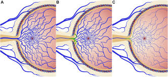
The primary cause of opticociliary shunt vessels is chronic obstruction or increased resistance in the central retinal vein, which impairs normal venous drainage from the eye. To bypass this obstruction, the body creates new venous pathways via the choroidal circulation. Causes include:
- Central Retinal Vein Occlusion (CRVO): A major cause, where a blockage in the central retinal vein leads to backup of blood flow and the development of collateral vessels.
- Chronic Papilledema: Long-term swelling of the optic disc (due to increased intracranial pressure) can lead to the formation of shunt vessels.
- Optic Nerve Sheath Meningioma: Tumors compressing the optic nerve can interfere with venous outflow.
- Glaucoma: Advanced glaucoma, especially with optic nerve damage, can lead to vascular remodeling and the appearance of shunt vessels.
- Orbital or Optic Nerve Tumors: Any mass effect in the orbit that compresses venous pathways can lead to the development of collaterals.
What Are the Symptoms of Opticociliary Shunt Vessels?
Opticociliary shunt vessels themselves do not cause symptoms, but they are often associated with underlying conditions that do. Patients may present with:
- Blurred Vision
- Gradual or Sudden Vision Loss
- Visual Field Defects
- Optic Disc Swelling
- Changes in Retinal Appearance
Because these vessels usually indicate an underlying issue, their discovery during an eye exam should prompt a thorough evaluation for related disease.
How Are Opticociliary Shunt Vessels Diagnosed?
These vessels are typically diagnosed during a comprehensive eye examination using specialized imaging tools:
- Fundus Examination: Using an ophthalmoscope or fundus camera, shunt vessels appear as dilated, tortuous veins crossing the optic disc.
- Optical Coherence Tomography (OCT): OCT can help assess optic nerve swelling and other structural changes.
- Fluorescein Angiography: This imaging test involves injecting dye into the bloodstream to visualize retinal and choroidal circulation. It can show the flow through the shunt vessels.
- Visual Field Testing: To assess any vision loss or field defects, especially if optic nerve compression is suspected.
- Neuroimaging (MRI or CT Scan): If optic nerve tumors or intracranial pressure are suspected, brain and orbital imaging may be required.
How Are Opticociliary Shunt Vessels Treated?

The shunt vessels themselves usually do not require direct treatment, but the underlying cause must be identified and managed. Treatment depends on the associated condition:
1. Managing Central Retinal Vein Occlusion (CRVO):
- Anti-VEGF Injections: To reduce macular edema and improve visual outcomes.
- Steroids: May be used in certain cases to reduce inflammation.
- Laser Therapy: Sometimes used for complications like neovascularization.
2. Treatment of Optic Nerve Tumors:
- Surgical Removal or Radiation Therapy: For tumors like optic nerve sheath meningiomas or orbital masses.
3. Control of Glaucoma:
- Eye Drops or Surgery: To reduce intraocular pressure and prevent further damage to the optic nerve.
4. Management of Papilledema:
- Treating the Underlying Cause of Increased Intracranial Pressure: Such as medication, lumbar puncture, or shunt surgery in cases of idiopathic intracranial hypertension.
Regular follow-up with an eye specialist is crucial to monitor the vessels and ensure that the underlying condition is appropriately managed.
What Is the Prognosis for Opticociliary Shunt Vessels?
The prognosis depends entirely on the underlying condition causing the formation of the vessels:
- If the underlying condition is mild or well-managed, visual outcomes may be stable.
- If opticociliary shunt vessels are a result of severe or chronic conditions like CRVO or tumors, vision may be permanently affected despite treatment.
- Early diagnosis and intervention in related diseases can improve outcomes and prevent progression.
When Should You See an Eye Specialist?
You should seek medical attention if you experience any of the following:
- Sudden or progressive vision loss
- Persistent visual field changes
- Signs of optic nerve swelling or eye pressure
- Any abnormal findings during a routine eye exam
Early detection of opticociliary shunt vessels can lead to diagnosis of serious underlying conditions before permanent vision damage occurs.
Can Opticociliary Shunt Vessels Be Prevented?
There is no way to directly prevent opticociliary shunt vessels since they are a secondary response to underlying pathology. However, you can reduce your risk by:
- Managing chronic conditions like hypertension, diabetes, and glaucoma
- Having regular eye exams, especially if you are at risk for retinal vascular diseases
- Promptly treating eye infections or trauma
- Monitoring for symptoms like vision loss or pressure in the eyes